15 Eylül 2025 Pazartesi
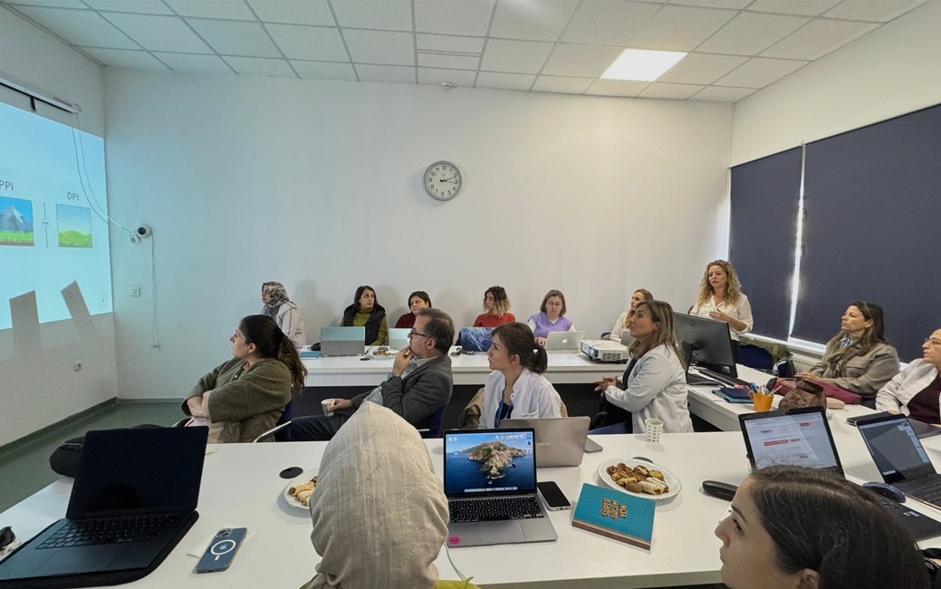
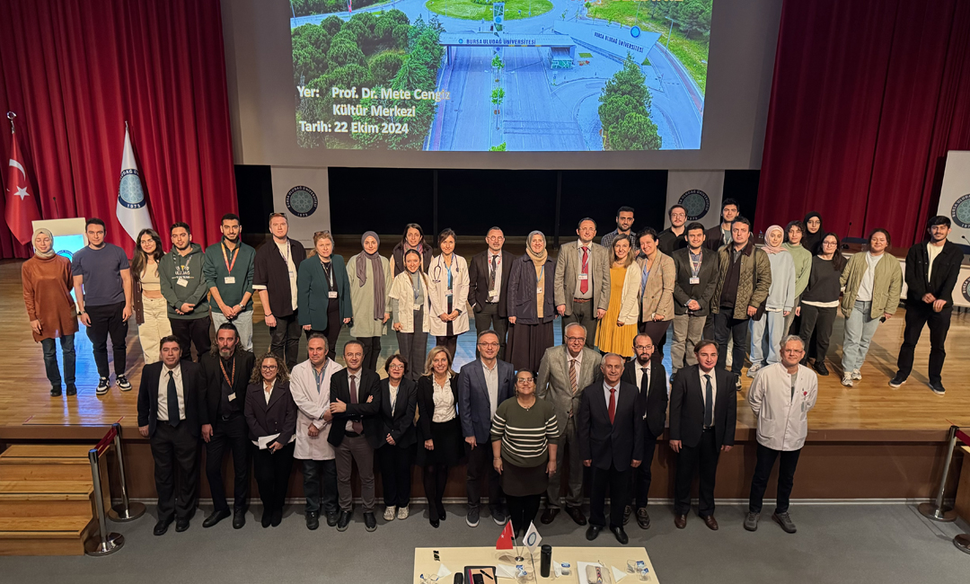
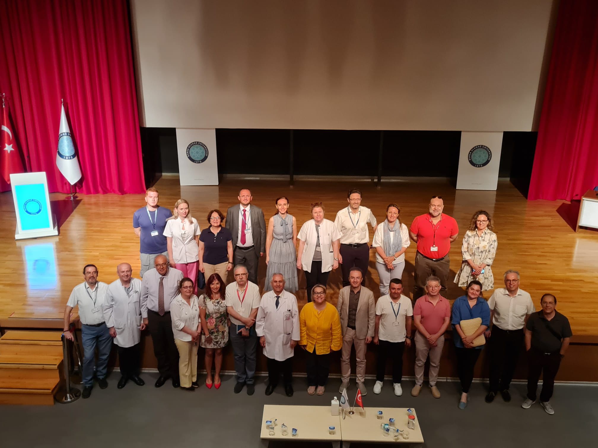
2025-2026 Eğitim Yılı Kliniğe Geçiş-Oryantasyon Günü Programı
Detaylı Bilgi
Deneme bonusu oyunculara risksiz deneme imkanı sunarken, Deneme bonusu veren siteler sayesinde yatırım yapmadan oyunları keşfedebilirsiniz. Papara bonusu veren siteler ise finansal işlemleri kolaylaştırarak ekstra avantaj sağlar. Doğru seçimle hem eğlenceyi artırabilir hem de kazancınızı güçlendirebilirsiniz.